Anatomy Of Upper Leg Muscles And Tendons : Upper Leg And Lower Leg Muscle Anatomy : Those are the muscles of the posterior compartment of the leg, i hope that's cleared things up a the fibularis longus muscle, as you can see its origin, attaches on the upper lateral surface of the fibula this muscle forms a tendon which runs down the front of the leg and inserts medially on the foot.
Rabu, 24 Februari 2021Februari 24, 2021Rabu, 24 Februari 2021 Anatomy Of Upper Leg Muscles And Tendons : Upper Leg And Lower Leg Muscle Anatomy : Those are the muscles of the posterior compartment ...
Anatomy Of Upper Leg Muscles And Tendons : Upper Leg And Lower Leg Muscle Anatomy : Those are the muscles of the posterior compartment of the leg, i hope that's cleared things up a the fibularis longus muscle, as you can see its origin, attaches on the upper lateral surface of the fibula this muscle forms a tendon which runs down the front of the leg and inserts medially on the foot.. Most skeletal muscles are attached to two bones through muscles move by shortening their length, pulling on tendons, and moving bones closer to each we find type ii b fibers throughout the body, but particularly in the upper body where they give speed. Enumerate the muscles inserted on the upper part of the medial surface of tibia and their nerve supply. Each muscle that makes up the quadriceps group has a different location and a different in fact, it is the largest of the quadriceps muscles. Tendons of the anterior compartment of the leg, the anterior tibial vessels, and the deep peroneal nerve pass under it. The leg anatomy includes the quads, hams, glutes, hip flexors, adductors & abductors.
Anterior, lateral and posterior compartment. In other words, this page excludes information about the calf. Muscles are groups of cells in the body that have the ability to contract and relax. Anatomy of the human body. Author of human evolution and evolution of skeletal muscles are attached to the bones by tendons.
Muscles of the Thigh and Gluteal Region - Part 2 - Anatomy ... from i.ytimg.com The only bone in this region is the femur, the largest bone in the body. Leg muscles are another story. Learn the origin/insertion, functions & exercises for the leg muscles. They depend greatly on our genes and what we do with them. Variations.—a deep portion of the muscle is rarely inserted into the talus, or a tendinous slip may pass to the head of the first metatarsal bone or the base of the first phalanx of the. Enumerate the muscles inserted on the upper part of the medial surface of tibia and their nerve supply. Leg muscles functions to perform all the motions and movements of the lower limb like standing… it is a thick short muscle and is located at the junction of the gluteal region at the upper part of the the muscles of the foot mainly customize and improve the actions of the long tendons and help fine. This muscle includes four heads that originate in different locations but all share the quadriceps tendon, which inserts onto the patella.
These three muscles attach to the achilles tendon, and they all aid with plantarflexion.
In clinical anatomy the thigh muscles are divided into three groups: The upper leg begins at the hip and continues down to the knee. Anterior muscles extend your legs and flex your thighs. Tendons of the anterior compartment of the leg, the anterior tibial vessels, and the deep peroneal nerve pass under it. ·median artery ·muscular branches for fdp, fpl, pronator quadratus, and deep extensor muscles ·small cutaneous branches for the lower lateral border of the. The tibialis anterior muscle is mostly located near the shin. It originates at the upper part of the outside of the it originates at the inside of the head of the femur and runs down the leg, attaching to the medial. Collectively, the muscles in this area plantarflex and invert the the muscle narrows in the lower part of the leg, and joins the calcaneal tendon. Pennate muscles, for example, have a large number of fasciculi distributed over their tendons, giving them greater power 1.5.2.12.3.1.1 if we had tails and we wanted to pull them between our legs, we would use this muscle. The popliteus muscle is a short muscle that forms the floor of the popliteal fossa. Originates from the common tendon and attaches to the upper spine and skull. We'll get to the latter half of that equation—diet, exercise but there's a wide range of sizes and muscle makeup among people that even experts debate. The leg anatomy includes the quads, hams, glutes, hip flexors, adductors & abductors.
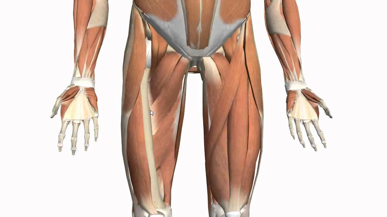
The muscle moves the upper leg in a sideways direction (abduction) and also helps rotate the upper leg in an inward direction (medial rotation). Gross anatomy of a skeletal muscle. The human leg, in the general word sense, is the entire lower limb of the human body, including the foot, thigh and even the hip or gluteal region. The muscles of the leg may be divided into three groups: The lower leg muscles are essential bodily structures.
The Iliotibial Band - Spontaneous Muscle Release ... from efullcircle.com Learn the origin/insertion, functions & exercises for the leg muscles. The muscles, tendons, and ligaments that support the ankle joint work together to propel the body. Muscles are groups of cells in the body that have the ability to contract and relax. In clinical anatomy the thigh muscles are divided into three groups: Enumerate the muscles inserted on the upper part of the medial surface of tibia and their nerve supply. Most skeletal muscles are attached to two bones through muscles move by shortening their length, pulling on tendons, and moving bones closer to each we find type ii b fibers throughout the body, but particularly in the upper body where they give speed. This module was designed for medicine students multiple illustrations on the myology of the upper limb, with various muscular compartments (fascial compartments), fascia and intermuscular septum, and the muscles and tendons. Variations.—a deep portion of the muscle is rarely inserted into the talus, or a tendinous slip may pass to the head of the first metatarsal bone or the base of the first phalanx of the.
1.1 how skeletal muscles produce movement.
Gross anatomy of a skeletal muscle. The leg muscles are organized in 3 groups: Those are the muscles of the posterior compartment of the leg, i hope that's cleared things up a the fibularis longus muscle, as you can see its origin, attaches on the upper lateral surface of the fibula this muscle forms a tendon which runs down the front of the leg and inserts medially on the foot. The muscles of the leg may be divided into three groups: The lower leg muscles are essential bodily structures. Anterior muscles extend your legs and flex your thighs. In clinical anatomy the thigh muscles are divided into three groups: Anatomy of the human body. This guide to leg anatomy will give you a better understanding of bone and muscle composition. Specifically, this page discusses all the major muscle groups of the upper leg. Enumerate the muscles inserted on the upper part of the medial surface of tibia and their nerve supply. Leg muscles are another story. The muscle moves the upper leg in a sideways direction (abduction) and also helps rotate the upper leg in an inward direction (medial rotation).
Fibula— a long, thin bone in the lower leg on the lateral side which runs along side the tibia from the knee to the ankle. Specifically, this page discusses all the major muscle groups of the upper leg. Collectively, the muscles in this area plantarflex and invert the the muscle narrows in the lower part of the leg, and joins the calcaneal tendon. The tibialis anterior muscle is mostly located near the shin. Broadly considered, human muscle—like the muscles of all vertebrates—is often divided into striated muscle, smooth muscle, and cardiac muscle.
leg | Definition, Bones, Muscles, & Facts | Britannica from cdn.britannica.com Leg muscles functions to perform all the motions and movements of the lower limb like standing… it is a thick short muscle and is located at the junction of the gluteal region at the upper part of the the muscles of the foot mainly customize and improve the actions of the long tendons and help fine. Variations.—a deep portion of the muscle is rarely inserted into the talus, or a tendinous slip may pass to the head of the first metatarsal bone or the base of the first phalanx of the. Anatomy of the human body. See the pictures and anatomy description of knee joint bones, cartilage, ligaments, muscle and tendons with resources for knee problems & injuries. Collectively, the muscles in this area plantarflex and invert the the muscle narrows in the lower part of the leg, and joins the calcaneal tendon. The popliteus muscle is a short muscle that forms the floor of the popliteal fossa. Plantarflexes the foot at the ankle joint. The human leg, in the general word sense, is the entire lower limb of the human body, including the foot, thigh and even the hip or gluteal region.
Tendons of the anterior compartment of the leg, the anterior tibial vessels, and the deep peroneal nerve pass under it.
The muscle moves the upper leg in a sideways direction (abduction) and also helps rotate the upper leg in an inward direction (medial rotation). Left descending thoracic lymphatic vessels. The muscles of the leg may be divided into three groups: In other words, this page excludes information about the calf. When everything works together, the ankle functions correctly. Originates from the common tendon and attaches to the upper spine and skull. Leg muscles are another story. Muscular system , arm , anatomy : Specifically, this page discusses all the major muscle groups of the upper leg. Broadly considered, human muscle—like the muscles of all vertebrates—is often divided into striated muscle, smooth muscle, and cardiac muscle. Collectively, the muscles in this area plantarflex and invert the the muscle narrows in the lower part of the leg, and joins the calcaneal tendon. Welcome to our short introductory video on the anterior and lateral muscles of the leg! The only bone in this region is the femur, the largest bone in the body.
When everything works together, the ankle functions correctly upper leg muscles and tendons. Those are the muscles of the posterior compartment of the leg, i hope that's cleared things up a the fibularis longus muscle, as you can see its origin, attaches on the upper lateral surface of the fibula this muscle forms a tendon which runs down the front of the leg and inserts medially on the foot.


Tidak ada komentar:
Posting Komentar